
Ultrasound

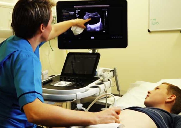
Ultrasound employs technology that sends sound waves into the body, recording the reflection. Ultrasound is also used for a detailed assessment of blood flow (‘Doppler ultrasound’) and for views of soft tissue and anatomical body structures.

Ultrasound is safe, painless and the first line investigation of choice for gallbladder and gynaecological abnormalities.

The Siemens Acuson 300 ultrasound machine employed in Wexford Ultrasound is the latest ultrasound technology from one of the world’s foremost suppliers of medical imaging technology.
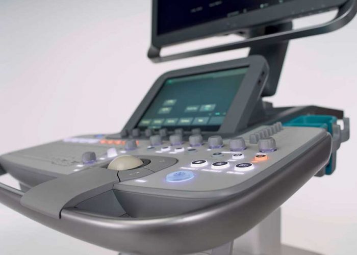
Siemans Acuson
Scan-5

Abdominal Ultrasound

Abdominal Ultrasound routinely involves the evaluation of the pancreas, midline abdominal vessels, liver, gallbladder, bile ducts, kidneys and spleen.

Fasting for 8 hours prior to imaging is required to allow full distension of the gallbladder and allow optimal evaluation.

Deep breaths in and holding the breath are required to allow visualisation of organs which lie behind the rib cage in the upper abdomen.

Examination takes approximately 15-20 minutes.

Pelvic Ultrasound

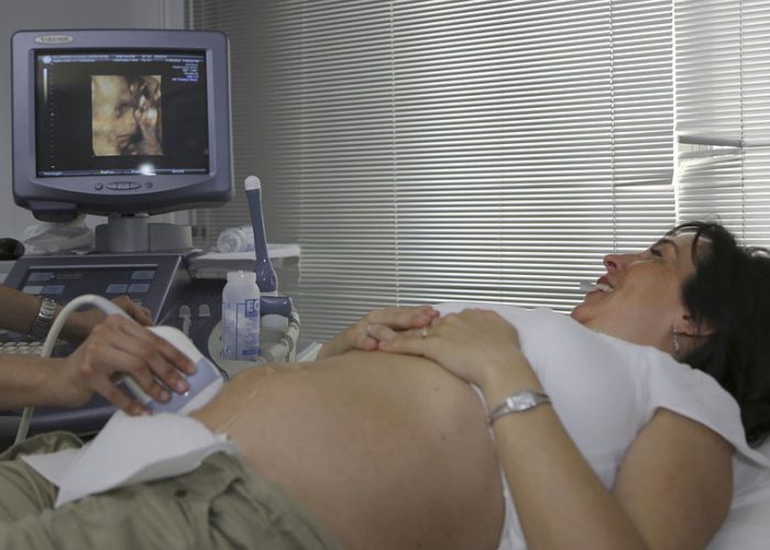
Pelvic Ultrasound can be either ‘transabdominal’ or ‘endovaginal’. Transabdominal scan is performed in the first instance, and requires a full urinary bladder to allow visualisation of the uterus (womb) and ovaries which lie behind the bladder.

Kidneys are also often imaged as part of the examination depending on the clinical presentation.

Each examination takes approximately 15-20 minutes.

Scan-2
Scan-3

Internal Examination (endovaginal)

In some circumstances, and depending on the clinical indications, an internal examination (endovaginal) is performed. This allows improved local visualisation of gynaecological anatomy, but is limited to structures immediately adjacent to the vagina. The patient is first asked to empty their bladder, then a narrow ultrasound probe is covered with a latex condom and the patient is asked to insert it into the vagina.

If deemed necessary endovaginal examinations can be organised offsite to be performed by female sonography staff in a controlled clinical environment.

Small parts Ultrasound

Small parts Ultrasound

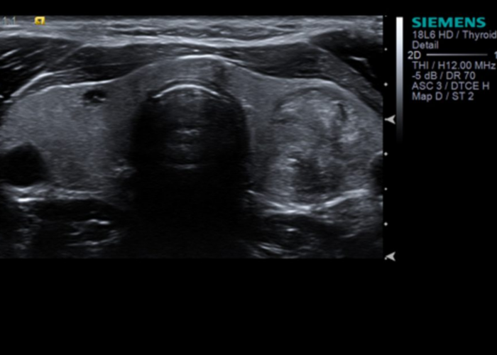
Small parts Ultrasound includes imaging of superficial structures such as the thyroid gland in the neck, testes, lymph glands and muscular and fatty layers beneath the skin.

These investigations do not require any specific preparation.

Doppler